After prostate cancer and bladder tumours, kidney cancer is the third most common malignant urological tumour. In Slovakia, approximately 600 new cases of kidney cancer are diagnosed every year. The incidence of kidney cancer is rising worldwide, especially in developed countries.
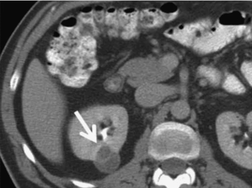
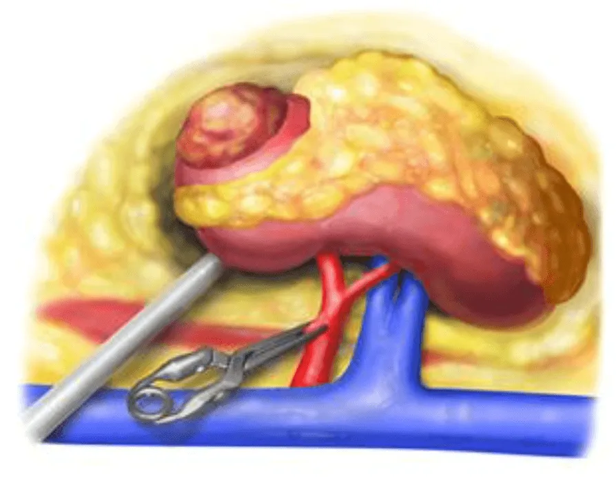
Thanks to the availability of modern imaging methods such as ultrasound, CT or MRI, kidney tumours are increasingly diagnosed at an early stage of the disease, that is, before any clinical symptoms appear. If kidney cancer already causes clinical symptoms /flank pain, blood in the urine, a palpable tumour/, it is a sign of advanced disease. Fortunately, today the vast majority of patients come to our department in the first stage of the disease. At this stage the disease is fully curable, and the treatment is surgery. If the kidney tumour is suitably located, it is not necessary to remove the whole kidney to cure the patient. It is enough to remove just the tumour with a small margin of healthy parenchyma. The patient therefore keeps practically the whole kidney, which is a huge advantage. In patients with a functionally or anatomically solitary kidney, radical removal of the whole kidney would lead to haemodialysis, with all its negative consequences for quality of life.
Robotic-assisted partial nephrectomy is performed with the da Vinci robotic system. At F. D. Roosevelt University Hospital we work with the da Vinci X system from Intuitive Surgical.
The operation is performed under general anaesthesia and takes approximately 1.5–2 hours. It is performed through access ports into the abdominal cavity (trocars), through which special instruments are inserted that the surgeon uses to carry out the whole procedure. The operation requires two experienced surgeons, one at the console and the other at the patient's side. The assistant helps with dissection and haemostasis, changes instruments, introduces sutures and clips and applies the vascular clamp.
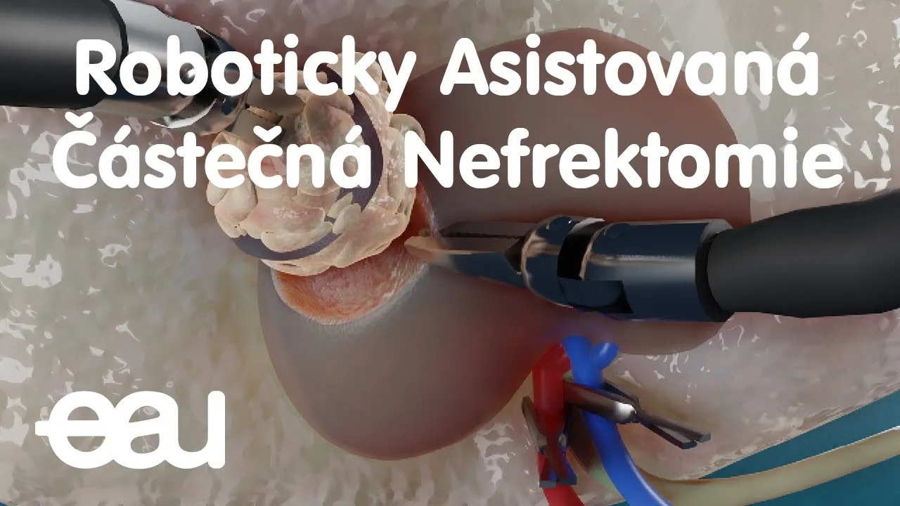
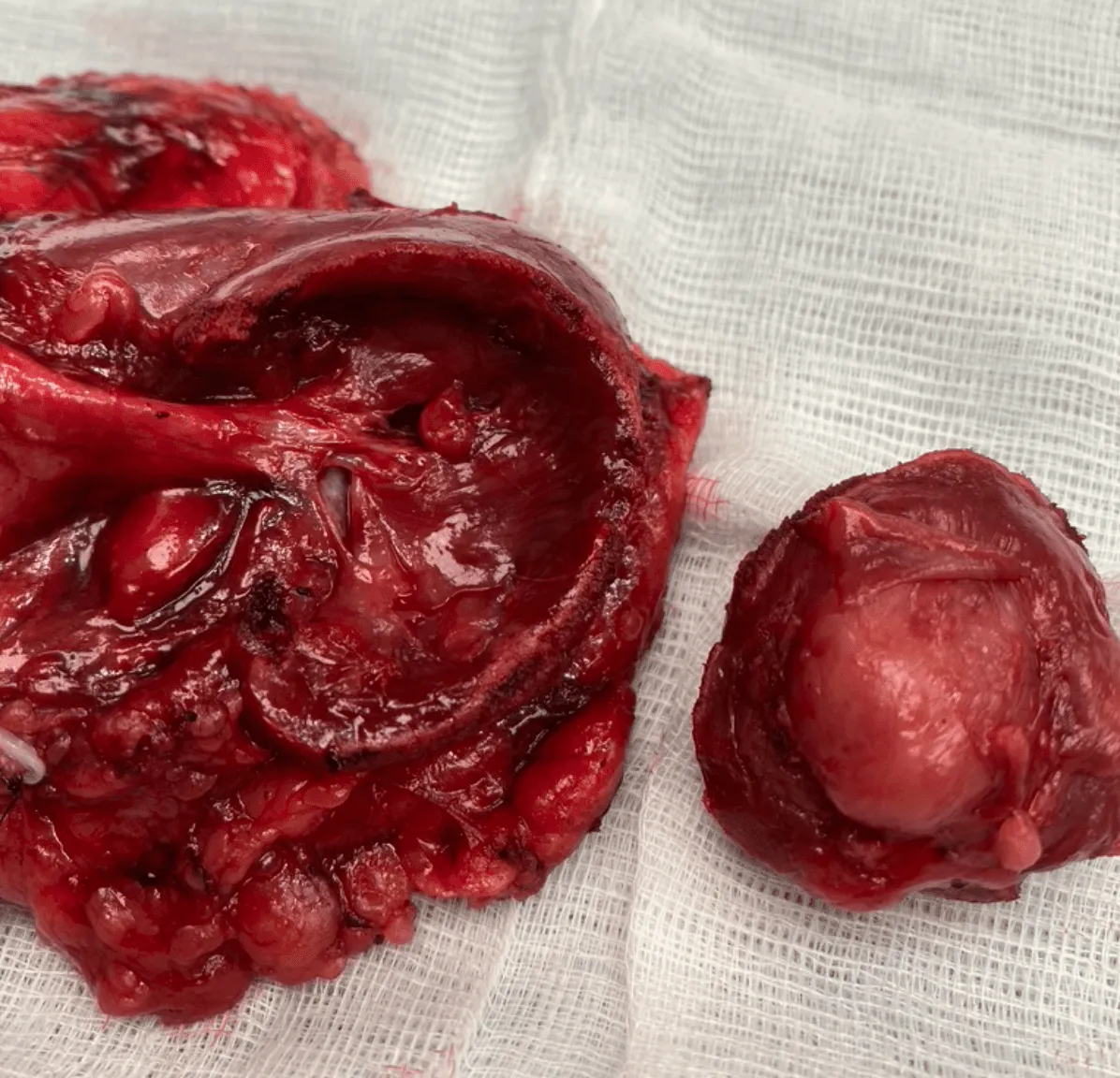
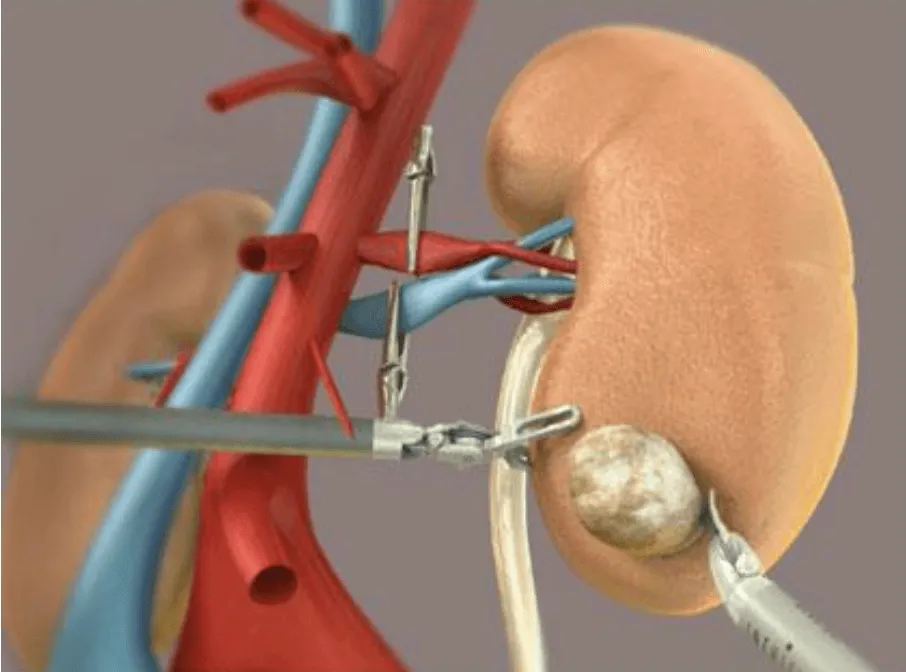
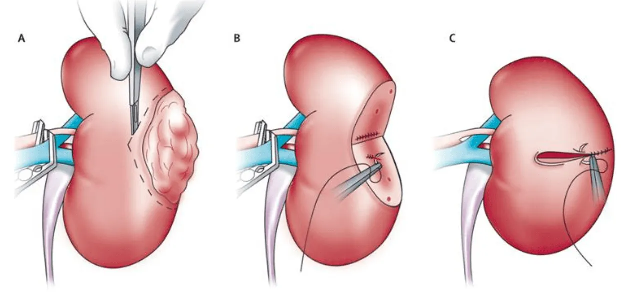
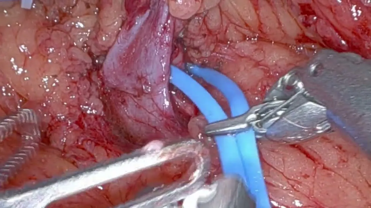
The patient is placed in the lumbotomy (flank) position. After the ports are inserted, docking follows – connecting the ports to the robot's arms. The extent of kidney mobilisation depends on the location of the tumour. In most cases extensive mobilisation of the kidney is not needed. This is followed by stopping blood flow through the kidney /so-called warm ischaemia/ by applying a vascular clamp, resection of the tumour and suturing of the tumour bed. After suturing, we check haemostasis and place a drain next to the kidney. The tumour removed from the kidney is taken out of the body in a special plastic bag through a small skin incision. The end result is 5 small scars on the abdominal wall.
Recording of a real operation:
Perhaps the greatest advantage of robotic partial resection over open surgery is the incomparably smaller incision. This of course means significantly less pain and faster recovery. While after open surgery the patient has a scar 20–25 cm long, robotic surgery leaves only a few tiny mini-incisions.
Safety and good results have been repeatedly demonstrated by several studies. Where it is technically possible to preserve the kidney, robotic-assisted kidney resection is a safe minimally invasive method, and at our department it is already the gold standard.
Further benefits:
- The most advanced surgical technique
- A precise procedure with a short warm ischaemia time
- Low blood loss
- Fewer post-operative complications
- Excellent oncological results
- A short hospital stay
- Cosmetic result
Before the procedure you need to undergo a thorough examination and a consultation with a urologist, who will assess your diagnosis, health, expectations and treatment options. The urologist will explain the course, risks and benefits of robotic-assisted partial nephrectomy and answer your questions. You will also be told how to prepare for the procedure, what to expect afterwards and how to look after yourself during recovery.
If you are interested in robotic-assisted partial nephrectomy, talk to your urologist and find out whether this treatment option for kidney cancer is suitable for you. Robotic-assisted partial nephrectomy is a modern and oncologically safe method that can improve your quality of life and prognosis.

For more information about robotic-assisted partial nephrectomy, see the following sources:
- robotika.fnspfdr.sk/oddelenia/o-robotickej-chirurgii-v-urologii/operacia-oblicky/
- patients.uroweb.org/cz/zhoubne-nadorove-onemocneni-ledvin/
Gallery